How is an X-ray obtained?
Despite more advanced imaging (i.e., MRI and CT) plain film X-rays are still essential for the diagnosis of many spine conditions. To produce an x-ray image, a generator (part of an x-ray machine) produces a beam of X-rays which is transmitted thru the portion of the body under evaluation. Some X-rays are absorbed more than others depending on the density and composition of the material being scanned (i.e., bone is denser than muscle). The X-rays that are not absorbed are captured on X-ray sensitive film. This creates an image which can be viewed.
Why are X-rays needed?
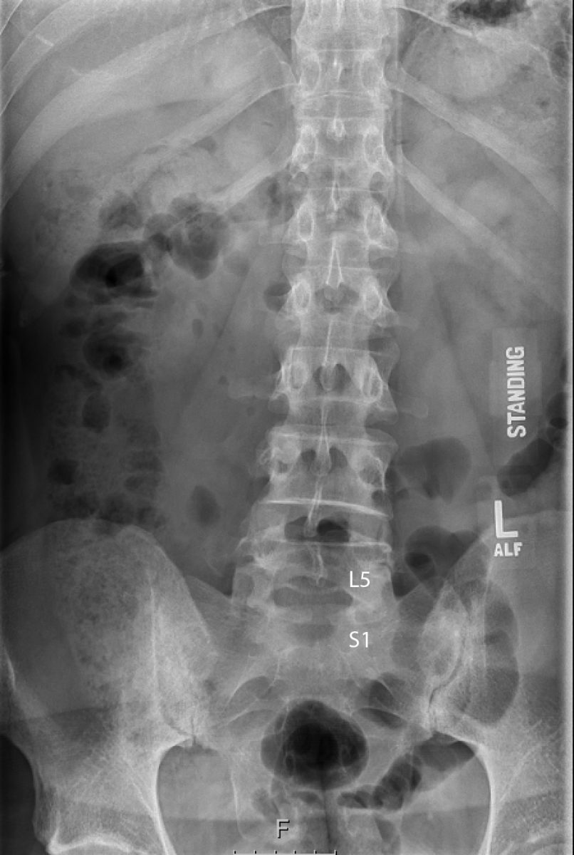
Some patients question why X-rays are necessary with the development of newer technologies such as CT and MRI. The advantage of x-rays is that they allow us to view the overall structure of one’s spine when they are standing. Abnormal curves (i.e., scoliosis and kyphosis) often improve when lying down. Therefore, the overall structure of the spine can be underappreciated with a test performed when a patient is lying on their back (i.e., MRI, CT). In addition, the diagnosis of a spondylolisthesis (when one vertebra slips forward with respect to another) may not be appreciated on a CT or MRI since these slips often reduce when a patient lies on their back. A CT and MRI are static tests since they show us what the spine looks when lying down. Plain X-rays are dynamic since we can take X-rays with the patient in different positions. This often includes a picture from the front and side of the patient when they are standing as well as leaning forwards and backwards while standing. This provides a lot of information including the overall alignment of the spine and abnormal curves (i.e., scoliosis and kyphosis).